CASE REPORT
Pseudoaneurysm of the anterior tibial artery following arthrodesis on a background of ankle joint tuberculosis
Veeralakshmanan P,1 Al-Saadi N,1 Popplewell M,1 Garnham A,1 Shalan A,1 Wall M1
Abstract
A 65-year-old man presented with a 6-week history of worsening right anterior ankle swelling following an ankle arthrodesis. Examination revealed a warm pulsatile 8 cm swelling on the dorsum of the foot. Ultrasound doppler and computed tomography angiography revealed an 8 cm pseudoaneurysm arising from the anterior tibial artery. Fluoroscopic-guided embolisation successfully treated the pseudoaneurysm. Pseudoaneurysms following foot and ankle surgery are a rare but documented complication. Treatment options include conservative management, endovascular interventions or surgical correction. This case highlights that awareness of blood supply variations in the foot and ankle is essential.
Case presentation
A 65-year-old man presented to the emergency department with a 6-week history of increasing right ankle swelling following elective right ankle arthroscopy (via right ankle anteromedial and anterolateral ports) and arthrodesis on a background of successfully treated ankle joint tuberculosis. The joint was fused in the appropriate position using cannulated screws. At the end of the procedure, an inadvertent breach of the anterior tibial cortex was noted but no specific action was required at that point and a plaster cast was applied with the foot in the plantigrade position. Following removal of the cast at 6 weeks, the right ankle was tender and swollen and the patient was unable to weight bear. He was on a prophylactic dose of low molecular weight heparin following the operation.
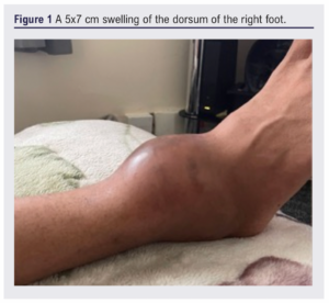
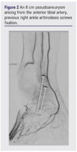
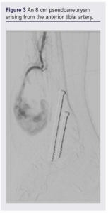
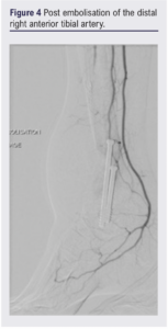
On examination a warm, pulsatile 5×7cm swelling was noted on the dorsum of the foot (Figure 1). The dorsalis pedis pulse was not palpable, but biphasic doppler signals were present. The rest of the pulse complexes and the contralateral side were unremarkable. Following an initial duplex scan, a computed tomography angiogram (CTA) confirmed an 8 cm anterior tibial artery pseudoaneurysm anterior to the surgical fixation site (Figures 2 and 3), which was subsequently embolised using fibred coils of 5×70 mm and 33×70 mm (Figure 4). Post-embolisation ultrasound confirmed thrombosis of the pseudoaneurysm. In the presence of the metal implants from the arthrodesis and the treatment coils, with no local or systemic signs of infection, it was elected not to evacuate the haematoma. Two days post embolisation the patient was discharged with thromboembolism deterrent stockings. At the 6-week follow-up there was a substantial reduction in anterior ankle swelling and he was able to weight bear and mobilise.
Discussion
Pseudoaneurysms are a rare complication following foot or ankle surgery with only four previously reported cases, two following intramedullary nailing for tibial fractures1,2 and two following ankle arthrodesis.3,4 Variation in arterial anatomy around the ankle is not uncommon, with changes in the course of the anterior tibial and peroneal arteries reported in 5% of cadavers.5 Appreciation of this anatomical variability is important when performing orthopaedic surgery and evaluating postoperative complications.
Pseudoaneurysm management varies with site and unit expertise. Options include conservative management, external ultrasound-guided compression,6 endovascular or surgical correction. Endovascular treatment options include coil embolisation, ultrasound-guided thrombin injection or stent insertion.7,8 Surgical management involves proximal and distal arterial control, haematoma evacuation (important if the pseudoaneurysm is associated with pressure effects) and arterial defect repair using direct closure, vein patching or interposition graft.
This case report of delayed pseudoaneurysm formation complicating orthopaedic ankle surgery highlights the importance of a heightened level of suspicion to prevent missed or delayed diagnoses. We encourage surgeons to remain vigilant for pseudoaneurysms when confronted with non-resolving haematomas including a low threshold for further investigations to prevent life-/limb-threatening complications.
Article DOI:
Journal Reference:
J.Vasc.Soc.G.B.Irel. 2024;3(2):108-110
Publication date:
February 29, 2024
Author Affiliations:
1. Vascular Surgery Department, Russells Hall Hospital, Black Country Vascular Network, West Midlands, UK
Corresponding author:
Pushpa Veeralakshmanan Vascular Surgery Department, Black Country Vascular Network, West Midlands DY1 2HQ, UK Email: p.veeralakshmanan @nhs.net


")

")

