ORIGINAL RESEARCH
Carotid web versus atherosclerotic carotid disease in surgical practice: a single-centre cohort of 579 carotid endarterectomies (2019–2024)
Binati FC, Kam M, Buckland J, Whitmore A, Howard DPJ
Plain English Summary
Why we undertook the work: A carotid web is a small shelf-like flap of tissue inside the carotid artery, the main blood vessel in the neck that supplies the brain. It is not caused by the fatty build-up (atherosclerosis) that accounts for most strokes, and it is often missed on standard ultrasound scans. Despite being rare, it can cause severe stroke, particularly in younger people. We wanted to understand how patients with a carotid web differ from those with the more common atherosclerotic type of carotid disease, and what happens to them before and after surgery.
What we did: We reviewed all 579 carotid artery operations performed at our hospital between 2019 and 2024. Every patient was assessed using detailed scan imaging and discussed by a specialist team before surgery. We identified 25 patients who had surgery for a carotid web and compared them in detail with the 554 patients who had surgery for atherosclerotic disease. We looked at age, sex, risk factors, scan findings, stroke severity and outcomes up to one year after surgery.
What we found: Carotid web patients were much younger (average age 53 vs 73 years) and more often female (56% vs 25%). They had fewer traditional risk factors such as high blood pressure or heart disease. Crucially, their standard neck ultrasound scan frequently appeared near-normal, even though they had suffered severe strokes – over half (52%) required emergency clot-busting treatment or mechanical clot removal before their operation compared with only 8% of atherosclerotic patients. Despite the severity of their initial presentation, patients who underwent surgery had no strokes in the first year after their operation.
What this means: Carotid webs can cause devastating strokes in young people who appear healthy on routine scans. They will be missed unless doctors specifically look for them using the right type of CT scan with detailed image reconstruction. We recommend that all young stroke patients should have a CT scan of their neck arteries as a matter of routine, and that imaging should include specific views designed to detect a carotid web. If one is found, urgent referral to a vascular surgeon should be made and prompt surgery considered.
Abstract
Background: Carotid web (CaW) is a shelf‑like, non‑atherosclerotic intimal lesion –histologically consistent with intimal fibroplasia – implicated in embolic stroke in younger, often female, patients. Comparative demographic, intervention and outcome data versus atherosclerotic disease are limited.
Methods: A single-centre study of consecutive carotid endarterectomies (CEAs) was performed for symptomatic carotid disease in 2019–2024, with follow-up to 2026. Cases were prospectively defined as CaW or atherosclerotic using multimodal imaging, operative reports and neurovascular multidisciplinary team assessment. We compared demographics, risk factors, lesion laterality/stenosis, clinical presentation (including National Institutes of Health Stroke Scale (NIHSS) score), emergency neurointervention prior to CEA and waiting period recurrence.
Results: Twenty-five of 579 CEAs (4.3%) were for CaW and 554 (95.7%) were for atherosclerosis. CaW patients were younger (53.3±11.9 years vs 73.0±10.0 years; p<0.001) and more often female (56.0% vs 25.1%; p=0.002), with fewer risk factors (hypertension 32.0% vs 70.8%, p<0.001; ischaemic heart disease 4.0% vs 25.8%, p=0.053; atrial fibrillation 0% vs 14.4%, p=0.044). CaW was predominantly unilateral (88.0% vs 46%, p<0.001) and frequently showed <50% duplex stenosis on the affected side (60.0% vs 0.2%, p<0.001). For CaW, presentation with stroke was more common (68.0% vs 43%, p=0.02) and the NIHSS score was higher (11.3±4.2 vs 6.0±3.9; p=0.009). Emergency thrombolysis and/or thrombectomy on initial presentation was also more frequent (52.0% for CaW (13/25) vs 7.9% for atherosclerotic disease (44/554), p<0.001). There was no significant difference regarding recurrent stroke whilst awaiting intervention (8.0% vs 2.2%, p=0.09) or postoperative stroke risk at 1 year follow-up (0% vs 2.1%, p=0.11).
Conclusion: CaW is an increasingly recognised cause of stroke in young patients. It represents a distinct embolic-prone non‑atherosclerotic pathology often presenting with severe stroke despite minimal detectable stenosis on duplex imaging and often subtle features on cross-sectional imaging. Prompt recognition on CT angiography and expedited definitive treatment are recommended.
Introduction
The carotid web (CaW) is characterised by a non-atherosclerotic fibrous ridge that appears as a shelf-like intraluminal projection, typically along the posterior wall of the carotid bulb or proximal internal carotid artery, leading to turbulent flow.1–3 CaW disproportionately affects younger patients – often women – with few traditional cardiovascular risk factors.1,4,5 Duplex ultrasound may underestimate CaW, frequently reporting it as minor stenosis only or minor focal plaque at the carotid bifurcation. Targeted cross-sectional imaging with oblique multiplanar reformats improves detection of the thin posterior shelf and associated mural thrombus.4-6 To date, there are no CaW-specific randomised trials, and comparative evidence for intervention (carotid endarterectomy (CEA) versus carotid artery stenting) is limited to small observational cohorts.7,8 Much of the published literature comprises case reports and case series only.9,10
Contemporary guidelines (ESVS, 2023) acknowledge the existence of CaW and support intervention for CaW in patients with recent symptoms, but provide no information on patient demographics, accurate diagnosis or treatment options for asymptomatic webs.11 No previous comparative studies of epidemiological and surgical data with direct comparison to atherosclerotic carotid disease have been published. We aimed to characterise and compare demographics, imaging features, presentation/severity, need for emergency neurointervention prior to CEA and waiting period recurrence between patients with CaW and those with atherosclerotic CEA. To our knowledge, this is the largest surgical series directly comparing patients undergoing CEA for symptomatic CaW with a contemporaneous atherosclerotic CEA cohort.
Methods
A single-centre cohort study was performed at a tertiary vascular unit (2019–2024). Data were extracted from institutional electronic patient records, a prospective clinical vascular database system and a prospectively maintained acute carotid referral system. Follow-up was maintained until 31 January 2026.
All patients undergoing symptomatic CEA were included. Exclusions comprised carotid dissection, tumour involvement and redo procedures. All patients with suspected CaW underwent CT angiography (CTA) with oblique multiplanar reformats. Imaging was reviewed in the neurovascular multidisciplinary team (MDT) meeting, including assessment by experienced vascular interventional radiologists, vascular surgeons and a consultant neurologist. Confirmation of diagnosis was made intraoperatively by direct visualisation of a web-like lesion at the carotid bulb (Figure 1).

All patients undergoing CEA for CaW received dual antiplatelet therapy postoperatively (aspirin 75 mg and clopidogrel 75 mg daily for three weeks), followed by lifelong single antiplatelet therapy (aspirin 75 mg daily).
Collected variables included age, sex, diagnosis of hypertension, ischaemic heart disease and atrial fibrillation, presenting syndrome (transient ischemic attack (TIA), stroke or amaurosis fugax), National Institutes of Health Stroke Scale (NIHSS) score at presentation, degree of stenosis measured by duplex (NASCET criteria) on the operated side, laterality (unilateral or bilateral disease), need for emergency neurointervention on presentation (thrombolysis and/or mechanical thrombectomy) and recurrent cerebrovascular events while awaiting CEA.
Continuous variables are presented as mean±SD and compared using an independent-samples t-test or Mann–Whitney U test, as appropriate. Categorical variables are reported as counts (%) and compared using the χ² test or Fisher’s exact test. All tests were two-tailed and p<0.05 was considered statistically significant. Analyses were performed in Python (v3.11) using pandas and SciPy, and with SPSS (version 31.0, 2025; SPSS Inc, Chicago, IL, USA).
The work was prospectively registered as clinical audit, ID number 9667.
Results
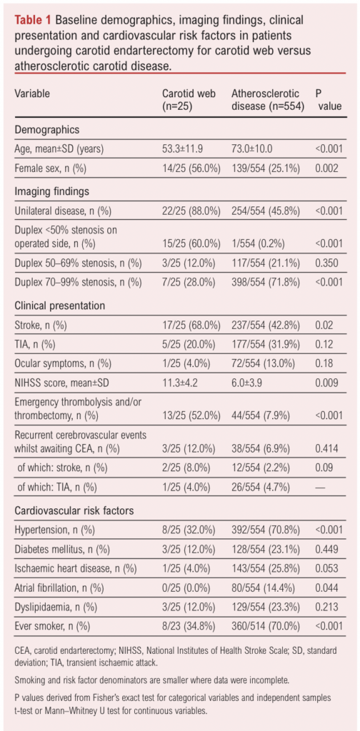
Of 579 CEAs, 25 (4.3%) were performed for CaW and 554 (95.7%) for atherosclerotic disease. Patients with CaW were significantly younger (53.3±11.9 years vs 73.0±10.0 years, p<0.001) and more frequently female (56.0% vs 25.1%, p=0.002). Compared with the atherosclerotic cohort, CaW patients had a lower prevalence of hypertension (32.0% vs 70.8%, p<0.001), ischaemic heart disease (4.0% vs 25.8%, p=0.053) and atrial fibrillation (0% vs 14.4%, p=0.044). Smoking history was more common in the atherosclerotic group (70.0% vs 34.8%, p<0.001). Baseline characteristics are summarised in Table 1.
Imaging findings
Operated-side duplex showed <50% stenosis in 60.0% of patients with CaW compared with 70–99% stenosis in 72% of atherosclerotic patients (p<0.001; Figure 2). CaW was predominantly unilateral (88.0% vs 46%, p<0.001).

Clinical presentation
At presentation, stroke was more common in the CaW group than in the atherosclerotic group (17/25 (68%) vs 237/554 (43%), p=0.02). Rates of TIA and other focal deficits were similar between groups (5/25 (20%) vs 177/554 (32%), p=0.12). Ocular symptoms tended towards being more common in the atherosclerotic group (72/554 (13%) vs 1/25 (4%), p=0.18). The mean NIHSS score was significantly higher in the CaW group (11.3±4.2 vs 6.0±3.9, p=0.009).
Emergency neurointervention
Emergency thrombolysis/thrombectomy was required in 52% of CaW patients compared with 8% of atherosclerotic patients (p<0.001).
Recurrent events
Recurrent cerebrovascular events whilst awaiting CEA were more common in the CaW group (12% vs 6.9%, p=0.414), comprising two strokes and one TIA in the CaW group. Stroke-specific recurrence was 8.0% vs 2.2% (p=0.09). The postoperative stroke rate at one year follow-up, inclusive of the perioperative period, was 0% in the CaW group versus 2.1% in the atherosclerotic group (p=0.11).
Discussion
In this large cohort study, CaW patients were younger, predominantly female and carried fewer classical vascular risk factors than atherosclerotic patients. CaW patients presented with more severe strokes (higher NIHSS score, more frequent need for emergency neurointervention) and often normal carotid duplex findings. This confirms data from case reports in the existing literature, where CaW has similarly been associated with younger age, female predominance and an embolic rather than stenotic clinical profile.1,4-6 Our results support prior observational work that characterise CaW as a distinct intimal arteriopathy completely separate from that of atherosclerotic carotid disease.1,4-6
Our study also confirms the presence of the hallmark clinical-radiological mismatch – namely, severe embolic events occurring in patients with often normal findings or minor stenosis on duplex imaging. The pathophysiological mechanism for this is flow stasis behind the posterior bulb intimal shelf and a recirculation pocket predisposing to thrombus formation. Consistent with prior case reports, we found that duplex can underestimate CaW, frequently reporting <50% stenosis using criteria developed to characterise atherosclerotic pathology (Figure 3). Cross-sectional imaging with multiplanar reformatting (and/or DSA where available) improves visualisation of the thin web and any associated mural thrombus.4-6,12 Current ESVS guidelines acknowledge CaW but offer no specific recommendations for accurate detection or management of asymptomatic webs, underscoring the persistent evidence gap.11 We propose a clinical service provision change whereby standardised checks for the presence of CaW are performed in all young stroke patients (defined as aged 60 years or under). This would require routine cross-sectional carotid imaging for all young stroke victims, and reporting standards to include multiplanar sagittal reconstruction to identify the presence or absence of CaW at the carotid bulb. If detected, urgent vascular surgery referral should be sought and expedited carotid surgery considered.

It is important to note that standard NASCET stenosis criteria, derived from atherosclerotic cohorts, should not be applied to guide the surgical indication in CaW. The mechanism of stroke in CaW is embolic rather than haemodynamic; 60% of CaW patients had <50% duplex stenosis yet 68% presented with stroke and 52% required emergency thrombolysis or thrombectomy. The surgical indication should be guided by the presence of symptoms and cross-sectional imaging confirmation of a web, not by the degree of luminal stenosis.13
Therapeutic implications
Symptomatic CaW carries a high risk of recurrent stroke on contemporary medical therapy alone.9,14 This risk is greatly reduced by urgent carotid intervention to remove the web and prevent further flow stasis and subsequent thrombus formation, and embolisation into the brain.7,8 We therefore support a clinical strategy of greater awareness, early diagnosis using the correct imaging format and expedited definitive treatment.
Whilst CEA directly removes the shelf and allows histological confirmation, carotid artery stenting offers a reasonable alternative in selected patients when surgical risk or anatomy favours endovascular treatment.7,9,10 Head-to-head comparative effectiveness data remain limited and justify further prospective studies to refine timing and modality.5,7,8
Strengths and limitations
The strengths of our work include the size of the surgical cohort and the presence of a large contemporaneous comparator group. This represents one of the largest comparative studies to date that directly compares patients undergoing CEA for symptomatic carotid webs versus atherosclerotic carotid disease. All cases underwent standardised preoperative imaging review (CTA with multiplanar reformats discussed in the vascular MDT) and intraoperative confirmation of CaW versus atherosclerotic plaque, reducing misclassification. Finally, we captured clinically meaningful stroke pathway prognostic markers, including requirement for pre-CEA thrombolysis/mechanical thrombectomy and neurological deficit at presentation.
Our work has limitations as it is a single-center design, has a modest CaW sample size (although significant considering the rarity of the disease) and the retrospective capture of some perioperative variables. As only patients undergoing carotid intervention were included and not all patients presenting with carotid territory cerebrovascular events, this selection bias requires acknowledgement when comparing these groups. Operative tissue was not routinely sent for histopathological analysis; however, intraoperative photographs were obtained in all 25 CaW cases to document the confirmed surgical diagnosis, providing a visual record of the characteristic shelf-like intimal lesion.
Research directions
We propose that future work on CaWs should include the following:
(i) Creation of an international registry to define natural history, including but not limited to stroke recurrence on best medical therapy and management of contralateral and/or asymptomatic disease.
(ii) Definition of standardised imaging criteria for the diagnosis of CaW as well as imaging markers for higher risk of embolization.
(iii) Prospective research to evaluate the role of expedited carotid surgery.
Hybrid PET imaging modalities including PET-CT and PET-MRI with targeted radiotracers such as 68Ga-FAPI, 18F-NaF and 18F-GP115-18 represent a promising avenue for future investigation. These techniques may provide functional information about lesion activity beyond that available on structural imaging alone. In particular, 18F-GP1, a platelet-targeted tracer, has demonstrated the ability to identify intraluminal thrombus in both the carotid artery and brain in acute stroke patients, including those with non-stenotic disease17,18 – a scenario directly analogous to CaW. Such approaches could potentially enable differentiation between active and quiescent carotid webs and refine patient selection for intervention. Their role in CaW management warrants prospective evaluation.
What this paper adds
There is currently no consensus on the optimal imaging modality for CaW diagnosis. Duplex ultrasound may identify an isoechoic or hypoechoic web structure with concurrent hypoechoic thrombus, but CaW can mimic ulcerated atherosclerotic lesions, limiting its diagnostic specificity.12 CTA remains the primary imaging technique of choice; it is safer and more accessible than MRI, and multiplanar reconstructions allow differentiation of CaW from localised dissections and aneurysms.19 MRI can be used as MRA or non-contrast time-of-flight (TOF) sequences, with T1-weighted imaging demonstrating high signal and T2-weighted imaging showing isointense or mildly elevated signal at the web. However, published data on MRI for CaW remain limited.12,19 Non-contrast MRI is particularly relevant in this population given the predominance of young women of childbearing age, in whom gadolinium-based contrast and radiation exposure should be minimised where possible.
CaW remains under-recognised in routine stroke work-up; on duplex ultrasound it is often undetectable or reported as mild stenosis only or minor focal plaque. However, patients with symptomatic CaWs are often young individuals presenting with major potentially life-changing strokes, who are at high risk of recurrent stroke if treated with contemporary medical therapy only.9,14 In our surgical cohort, patients undergoing CEA for CaW were younger and more frequently female, and they had substantially higher rates of emergency pre-CEA reperfusion procedures (intravenous thrombolysis and/or mechanical thrombectomy) compared with those treated for atherosclerotic disease (52% vs 8%). These findings support a service change in which cross-sectional carotid imaging is routinely performed for all young stroke victims, and reporting standards include sagittal reconstruction to identify the presence of CaW at the carotid bulb. If detected, urgent vascular surgery referral should be sought and expedited carotid surgery considered.
Conclusion
CaW is an increasingly recognised cause of stroke in young patients. It represents a distinct, embolic-prone, non-atherosclerotic pathology, often presenting with severe stroke despite minimal detectable stenosis on duplex imaging and often subtle features on cross-sectional imaging. Prompt recognition and expedited definitive treatment appear warranted. Multicentre data are needed to guide management, particularly for asymptomatic CaW disease.

Article DOI:
Journal Reference:
J.Vasc.Soc.G.B.Irel. 2026;Online ahead of publication
Publication date:
November 20, 2026
Author Affiliations:
Department of Vascular Surgery,
John Radcliffe Hospital,
Oxford University Hospitals NHS
Foundation Trust, Headington, Oxford, UK
Corresponding author:
Ms Fernanda Carolina Binati Department of Vascular Surgery, John Radcliffe Hospital, Headley Way, Headington, Oxford OX3 9DU, UK
Email: [email protected]