
CASE REPORT
Endovascular coil embolisation of radial artery pseudoaneurysm: a case report and literature review
Daysley H,1 Hamadneh M,2 Chikhhal R2
Abstract
Background: Radial artery pseudoaneurysm (RAP) is a rare but recognised complication of transradial percutaneous coronary intervention (PCI) with an incidence of 0.009–0.2%. Management depends on pseudoaneurysm size, morphology and patient factors. While conservative measures are often effective for small lesions, larger or symptomatic pseudoaneurysms may require surgical or endovascular intervention.
Case report: A 77-year-old woman developed a right radial artery pseudoaneurysm following transradial PCI. Initial duplex ultrasound demonstrated a 16 × 10 × 13 mm pseudoaneurysm, which was managed conservatively. Two weeks later she presented with worsening wrist pain, swelling and extensive bruising. Repeat ultrasound demonstrated enlargement to 45 × 21 × 29 mm with a 16 mm neck, making thrombin injection unsuitable. Owing to recent myocardial infarction and high anaesthetic risk, open surgical repair was considered inappropriate. A digital subtraction angiogram confirmed adequate ulnar collateral circulation. Following multidisciplinary discussion, endovascular coil embolisation of the radial artery was performed successfully using detachable Ruby coils. Post-procedural angiography and duplex ultrasound confirmed complete occlusion of the radial artery and pseudoaneurysm without evidence of hand ischaemia. The patient recovered without complications and was discharged four days later.
Conclusion: This case demonstrates that endovascular coil embolisation is a safe and effective treatment for enlarging radial artery pseudoaneurysms in carefully selected patients unsuitable for open surgery. Pre-procedural assessment of collateral hand perfusion and multidisciplinary decision-making are essential to achieve favourable outcomes.
Introduction
Radial artery pseudoaneurysms (RAP) are primarily caused by iatrogenic trauma. The transradial approach has become the preferred access route for cardiac catheterisation due to lower rates of bleeding and vascular morbidity compared with femoral access.1 RAP following percutaneous coronary intervention (PCI) are rare with incidences ranging from 0.009% to 0.2% of radial catheterisations, including PCI cases.1–4 Pseudoaneurysms occur due to a breach of the arterial wall with persistent communication between the lumen and surrounding soft tissue, often exacerbated by risk factors such as anticoagulation, multiple puncture attempts, incomplete haemostasis, advanced age and larger sheath sizes.
Management is guided by pseudoaneurysm size, symptoms and patient risk profile. Conservative measures such as stopping anticoagulation, ultrasound-guided compression and thrombin injection have been successfully employed, while surgical repair or endovascular techniques are often reserved for larger or refractory lesions.3 In this case report we describe the management of a RAP following PCI.
Case report
Preoperative assessment
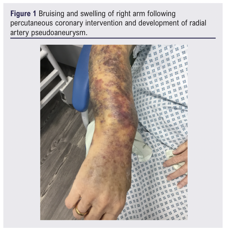
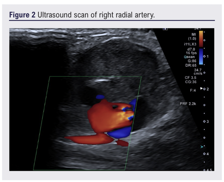
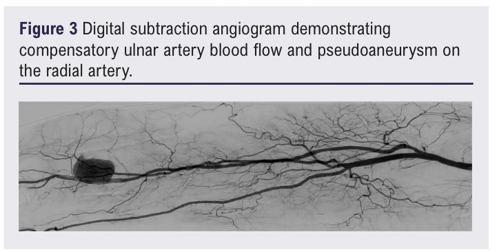
A 77-year-old woman presented with progressive right wrist swelling, pain, bruising and collapse. Two weeks earlier she had undergone PCI via a right radial approach following a myocardial infarction, which was complicated by RAP (16 × 10 × 13 mm) managed with ultrasound-guided compression. Comorbidities included atrial fibrillation and hypertension, managed with edoxaban, bisoprolol and ramipril. Examination revealed extensive right forearm and hand bruising, normal motor and sensory function, a palpable ulnar pulse and a warm well perfused hand with normal capillary refill time. There was a tender pulsatile 4 cm diameter mass over the radial artery at the wrist (Figure 1). A Modified Allen’s test confirmed a patent palmer arch and a peripheral duplex scan confirmed a RAP measuring 45 × 21 × 29 mm with a neck diameter of 16 mm (Figure 2). A digital subtraction angiogram confirmed the RAP and patency of both radial and ulnar arteries (Figure 3). The patient was discussed at the multidisciplinary meeting. Further ultrasound-guided compression and thrombin injection were precluded by previous failure, pain and RAP neck size. The patient was deemed a high-risk candidate for general anaesthesia due to recent cardiac events and highly unlikely to tolerate a surgical procedure under local anaesthetic. Endovascular options were discussed and embolisation was considered the most appropriate intervention option.
Endovascular management
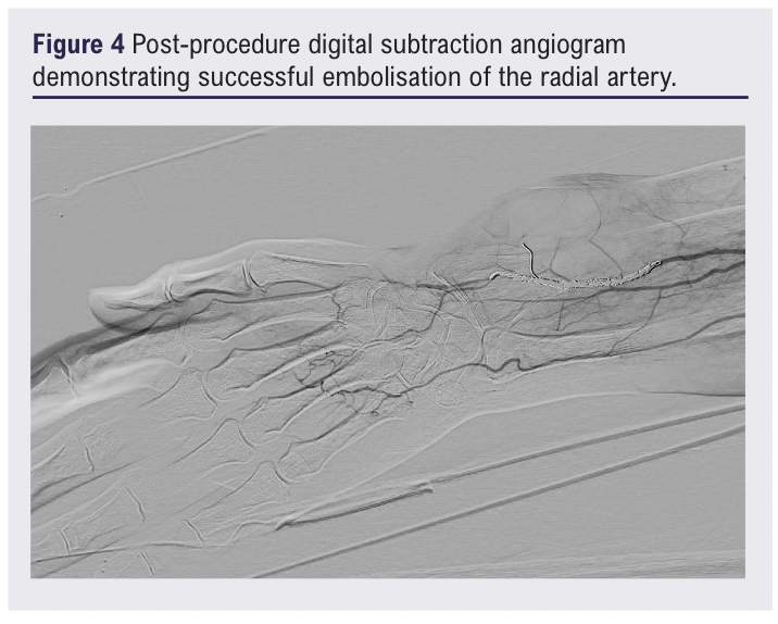
Forty-eight hours prior to the procedure the patient’s edoxaban was stopped and she was placed on prophylactic enoxaparin to minimise the risks associated with atrial fibrillation. Under local anaesthetic via 4F sheath right brachial artery access, right radial artery embolisation with successful exclusion of the RAP was achieved using five ruby coils (2 mm × 4 cm, 2 mm × 2 cm, 3 mm × 5 cm, 4 mm × 6 cm and 3 mm × 20 mm sizes) and one packing coil (15 cm) (Figure 4). Brachial access haemostasis was achieved with compression.
Postoperative management and outcomes
Postoperatively, the patient’s pain improved and the hand remained warm pink and well perfused with no neurological deficit. The ulnar pulse was palpable but there was no pulse in the distal radial artery or RAP. Duplex ultrasound confirmed successful distal radial artery embolisation and successful exclusion of the RAP. After 24 hours the patient was restarted on edoxaban and, following review by both the physiotherapy and occupation therapy teams, was discharged four days after the intervention. At 6-week follow-up the patient had minimal symptoms and the extent of the bruising and size of the RAP were reducing.
Discussion
RAP remains an uncommon complication following transradial coronary intervention with a reported incidence of 0.009–0.2%.1-4 A recent large systematic review analysing 75 published RAP cases noted that approximately 76% of patients developed symptoms or enlargement requiring active intervention, while only a minority were successfully managed conservatively.5 RAP recognised risk factors include multiple puncture attempts, larger sheath size, inadequate haemostasis, prolonged catheterisation, anticoagulation and advanced age.6,7 Our patient exhibited several of these factors.
Management is guided by pseudoaneurysm size, neck morphology, symptoms and patient comorbidity. Small (<2–3 cm) asymptomatic pseudoaneurysms may be observed, as spontaneous thrombosis has been reported.8 Ultrasound-guided compression remains an accepted first-line treatment; however, it is often painful, requires prolonged compression and is less effective in anticoagulated patients or wide-neck lesions. The recent systematic review reported a success rate of only 58.5% for compression therapy, with many patients requiring subsequent intervention because of persistent flow or interval enlargement.5 The failure of initial compression in our patient is therefore consistent with published experience.
Ultrasound-guided thrombin injection has become the preferred minimally invasive treatment for many iatrogenic peripheral pseudoaneurysms because of its high efficacy and low morbidity. Large series report technical success rates of 95–99%, with recurrence rates generally below 5%.9.10 However, treatment is dependent on favourable anatomy, particularly a relatively narrow neck that minimises the risk of thrombin entering the parent artery and causing distal embolisation. Wide-neck pseudoaneurysms, rapidly enlarging lesions and lesions with complex morphology are generally considered unsuitable for thrombin injection.10 In this case, enlargement from 16 mm to 45 mm with a wide neck following failed compression rendered thrombin injection inappropriate because of the published associated risks.
Open surgical repair has historically been regarded as the definitive treatment for symptomatic, enlarging or complicated RAPs. Reported surgical success rates exceed 95%, and recurrence is uncommon.5,11 Risks associated with surgery include wound infection, haematoma, sensory nerve injury, and the risks of regional or general anaesthesia. These considerations are particularly important in elderly patients with significant cardiovascular comorbidity. In our patient, recent myocardial infarction, ongoing anticoagulation, and poor tolerance of general anaesthesia or prolonged local anaesthetic substantially increased operative risk, making open repair less attractive.
Endovascular techniques provide an alternative for poor surgical candidates. Covered stent placement has been described with preservation of radial artery patency, but experience remains limited to isolated case reports.12 Deployment is technically challenging because of the small calibre and tortuosity of the radial artery, and long-term patency remains uncertain, particularly in elderly patients. The wide pseudoaneurysm neck and relatively small parent vessel in this case made durable stent exclusion unlikely and increased the risk of acute thrombosis or endoleak.
Coil embolisation is less frequently reported but represents an effective strategy when sacrifice of the radial artery is acceptable. Published experience is limited to case reports and small series; however, these reports consistently demonstrate technical success rates approaching 100%, durable pseudoaneurysm exclusion and very low complication rates when adequate collateral circulation is present.5,13,14 In the systematic review by Hajeh et al, all reported patients treated with percutaneous embolisation achieved successful exclusion of the pseudoaneurysm without hand ischaemia.5 Compared with covered stents, coil embolisation is technically simpler, avoids the need for long-term stent surveillance and eliminates the possibility of persistent sac perfusion. The principal prerequisite is confirmation that the ulnar artery and palmar arch can maintain hand perfusion. In our patient, angiography confirmed robust ulnar inflow and a complete palmar arch, permitting safe radial artery sacrifice without digital ischaemia. Adequate perfusion is confirmed angiographically by inserting a suitable sized occlusion balloon into the radial artery, then injecting contrast into the access site, documenting radial and ulnar patency, superficial and deep palmar arch, digital arterial filling and any anatomical variants.
An alternative endovascular strategy would have been selective coil embolisation of the pseudoaneurysm sac or neck while preserving radial artery patency. This approach offers the theoretical advantage of definitive pseudoaneurysm exclusion without sacrificing the radial artery and may therefore be preferable where the anatomy permits stable coil deployment. Recent case reports have demonstrated successful selective coil embolisation of radial artery pseudoaneurysms with preservation of the parent artery.13,14 The feasibility of this technique depends on the morphology of the pseudoaneurysm, as coils must be positioned securely within the sac or across the neck without protrusion into the parent vessel. In our patient, the relatively large 16 mm neck and rapidly enlarging 45 mm pseudoaneurysm would have increased the risk of coil migration or incomplete exclusion, making selective sac embolisation less predictable. Given the confirmed robust ulnar collateral circulation, intentional radial artery embolisation was therefore considered a more definitive approach.
An additional technical consideration was the use of brachial artery access. This provided a short direct route to the target vessel and allowed the procedure to be performed entirely under local anaesthesia. A recent systematic review of brachial artery access for endovascular interventions reported pooled complication rates of 4.8% for haematoma, 1.4% for haemorrhage, 1.1% for pseudoaneurysm, 0.6% for arterial thrombosis, 0.5% for neuropathy and 0.4% for limb ischaemia.15 In this case, haemostasis was achieved with manual compression alone and no access site complications occurred, supporting brachial access as a safe and practical option for selected upper limb interventions.
This report adds to the limited literature supporting endovascular coil embolisation for complex RAP after transradial PCI. Most published RAP cases continue to be managed surgically or with thrombin injection.5 Our case demonstrates that coil embolisation can provide definitive treatment after failed compression in a rapidly enlarging wide-neck pseudoaneurysm in a patient unsuitable for surgery, with preservation of hand perfusion and no neurological or access site complications.
A limitation of this case report is the lack of saved radiological imaging to support the findings of an intact palmar arch, despite that being documented and discussed in her case and management. However, these findings were supported by the clinical assessments including modified Allen’s test and postoperative pulses and neurovascular findings. A further limitation is the absence of long-term follow-up beyond the early postoperative period. Although immediate technical and clinical success was achieved, longer surveillance would be valuable to confirm complete pseudoaneurysm resolution and preservation of hand function.
Conclusion
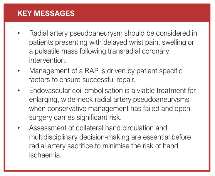
Radial artery pseudoaneurysm following transradial PCI remains an uncommon complication but should be considered in any patient presenting with delayed wrist swelling, pain or a pulsatile mass after catheterisation. Prompt duplex ultrasound facilitates diagnosis and guides management. This case illustrates that endovascular coil embolisation can provide definitive treatment when conservative management has failed and surgery carries excessive risk. Careful pre-procedural assessment of collateral hand circulation and multidisciplinary decision making are essential to optimise outcomes.
Article DOI:
Journal Reference:
J.Vasc.Soc.G.B.Irel. 2026;5(4):195-198
Publication date:
August 26, 2026
Author Affiliations:
1. Centre of Clinical Sciences, Hull York Medical School and Academic Vascular Surgical Unit, Hull, UK
2. Academic Vascular Surgical Unit, Hull, UK
Corresponding author:
Dr Hannah Daysley
Vascular Department,
Hull Royal Infirmary,
Anlaby Road,
Hull HU3 2JZ, UK
Email: [email protected]