
CASE REPORT
Splenic artery pseudoaneurysm presenting with upper gastrointestinal bleeding successfully treated by endovascular embolisation: a case report
Márquez JA,1 Llanos Luengas LV,2 Mendoza LF2
Abstract
Background: A splenic artery pseudoaneurysm is an uncommon vascular lesion accounting for less than 3% of splenic artery aneurysmal lesions, associated with a high risk of rupture (37–47%) and mortality rates up to 90%. Its clinical presentation is often non-specific and may include upper gastrointestinal bleeding, making early diagnosis crucial.
Case presentation: An 80-year-old man was admitted with acute melaena and severe anaemia. Emergency upper gastrointestinal endoscopy revealed an apparent ‘ulcerated subepithelial gastric lesion’ associated with active bleeding that could not be controlled with epinephrine injection. Computed tomography angiography showed a 42 mm vascular lesion arising from the splenic artery, irregular contour, absence of mural calcification, an eccentric configuration in close contact with the posterior gastric wall, without a clearly identifiable fistulous or epithelialised tract between the two structures, raising a high suspicion for a splenic artery pseudoaneurysm. Urgent angiography confirmed the diagnosis. Endovascular treatment was successfully performed with proximal and distal coil embolisation, achieving complete exclusion of the lesion. Follow-up endoscopy confirmed successful haemostasis and the patient was discharged four days later.
Conclusion: Splenic artery pseudoaneurysm can present with unexplained upper gastrointestinal bleeding. Early endovascular embolisation appears to be safe and effective.
Introduction
Splenic artery pseudoaneurysms are rare vascular lesions, accounting for less than 3% of splenic artery aneurysmal lesions, and are most commonly associated with pancreatitis, trauma and peptic ulcer disease.1 Clinical presentation is frequently non-specific, with abdominal pain and gastrointestinal bleeding being the most common manifestations. Early diagnosis using appropriate imaging modalities followed by prompt treatment is recommended to prevent rupture, which has been reported in 37–47% of cases and has an associated high mortality. Endovascular management, including embolisation or stent graft placement, has emerged as the preferred and effective minimally invasive therapeutic treatment strategy; however, potential complications including splenic infarction should be considered.2 We report an unusual case of an 80-year-old man with a splenic artery pseudoaneurysm presenting with upper gastrointestinal bleeding who was successfully treated with an endovascular approach.
Case presentation
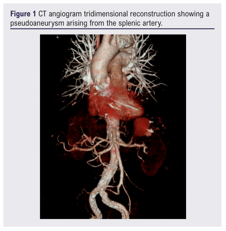
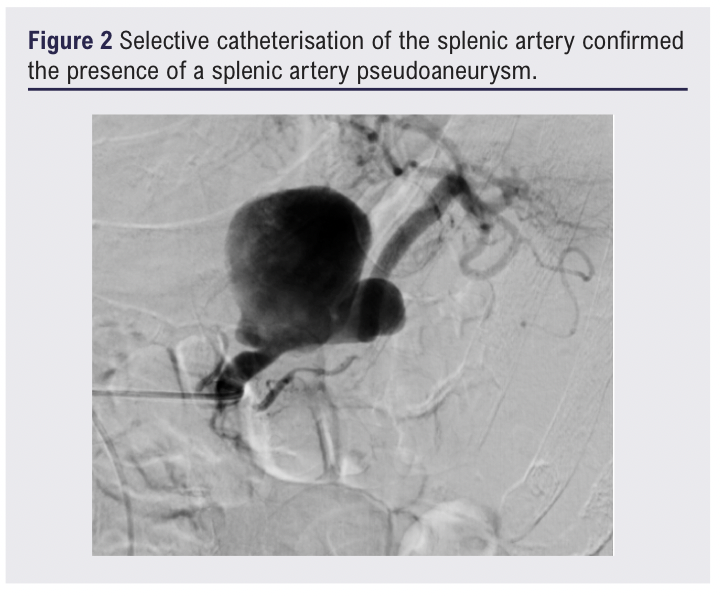
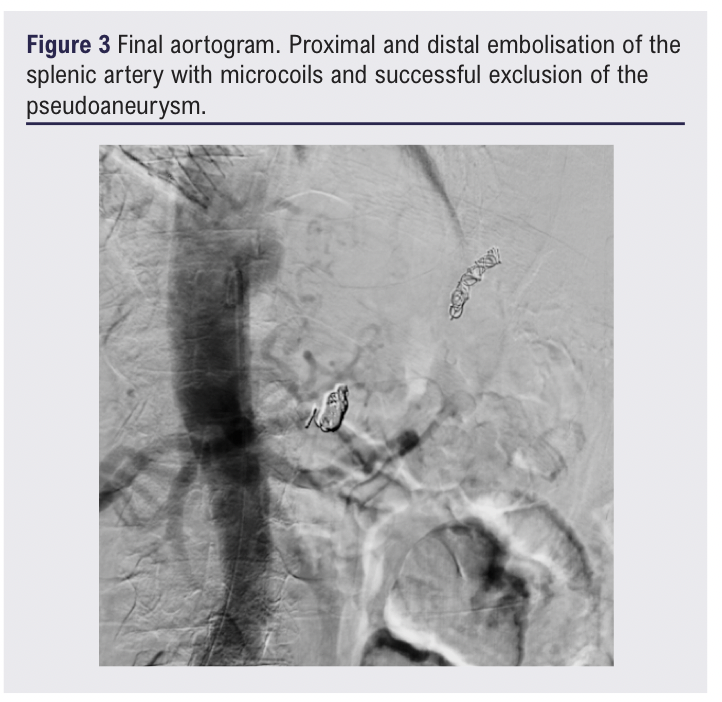
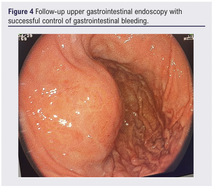
An 80-year-old man presented to the emergency room with upper gastrointestinal bleeding manifesting with sudden melaena. He denied abdominal pain or constitutional symptoms and had a history of hypertension, dyslipidaemia, right eye glaucoma and critical aortic valve stenosis with a prior transcatheter aortic valve replacement (TAVI) one year earlier. He denied tobacco or alcohol use. On admission he was haemodynamically stable but anaemic (haemoglobin 8.1 g/dL, haematocrit 25.4%). Emergency upper gastrointestinal endoscopy performed by the gastroenterology team revealed a bloody gastric residue and an apparent 5 cm subepithelial lesion, with superficial gastric mucosal ulceration along the greater curve with active bleeding, without a fistulous connection clearly identified. Endoscopic haemostasis was attempted with epinephrine injection but failed to achieve bleeding control. CT angiography (CTA) showed a well-defined vascular lesion arising from the splenic artery, measuring 38 × 39 × 42 mm (craniocaudal × anteroposterior × transverse dimensions) characterised by an irregular contour, absence of mural calcification, an eccentric configuration and intense arterial contrast enhancement (Figure 1). The lesion was in close contact with the posterior gastric wall, without a clearly identifiable fistulous or epithelialised tract between the two structures, raising a high suspicion for a splenic artery pseudoaneurysm. The patient required transfusion of two units of packed red blood cells. However, without an adequate clinical response and persistent upper gastrointestinal bleeding, urgent transfemoral visceral angiography and selective catheterisation of the splenic artery confirmed the presence of a splenic artery pseudoaneurysm (Figure 2). Endovascular treatment was successfully performed using sandwich embolisation with proximal (four microcoils) and distal (three microcoils) coil deployment, achieving complete exclusion of the pseudoaneurysm without procedural complications (Figure 3). The patient was admitted to the intensive care unit for close monitoring. Control upper gastrointestinal endoscopy showed adequate gastrointestinal bleeding control (Figure 4) and the patient was discharged home four days after the procedure.
Discussion
Splenic artery aneurysms are the most common visceral arterial aneurysms after aortic and common iliac artery aneurysms. True splenic artery aneurysms have a prevalence of less than 1%,3 and splenic artery pseudoaneurysms are even rarer. Frequent aetiologies of splenic artery pseudoaneurysms include pancreatitis and direct trauma to the splenic artery. In the present case we speculate that the pseudoaneurysm may have resulted from inadvertent guidewire- or catheter-related injury to the splenic artery during the patient’s previous TAVI. Splenic artery pseudoaneurysms have a variable clinical presentation, ranging from mild abdominal pain to life-threatening rupture or gastrointestinal fistulation. The associated risk of rupture may be as high as 47%,4 so early clinical suspicion, accurate diagnosis and prompt treatment if appropriate is essential. CTA is the most accessible non-invasive and preferred imaging modality for diagnosis. However, in the setting of active haemorrhage, catheter angiography remains the diagnostic and therapeutic gold standard. Different endovascular interventions can be considered including coil embolisation (coil trapping, isolation or sandwich technique) to prevent continued sac pressurisation from the collateral circulation, or covered stent placement.3 Splenic infarction remains one of its most common complications. Despite the rich collateral blood supply to the spleen, partial splenic infarction has been reported in up to 40% of patients following embolisation.5 Level 2/3 post-procedural monitoring is therefore recommended.
Conclusions
Splenic artery pseudoaneurysm is a rare but potentially fatal condition due to its significant risk of rupture. Clinical manifestations are often non-specific and a high index of suspicion is crucial, especially in patients with gastrointestinal bleeding. CTA and intra-arterial angiography play a key role in the diagnosis and treatment. This case highlights the effectiveness of endovascular embolisation allowing definitive haemorrhage control with excellent outcomes. Early recognition and timely intervention remain critical to reducing morbidity and mortality.
Article DOI:
Journal Reference:
J.Vasc.Soc.G.B.Irel. 2026;5(4):192-194
Publication date:
August 11, 2026
Author Affiliations:
1. Vascular and Endovascular Surgeon, Sociedad de Cirugía de Bogotá Hospital de San José, Bogotá, Colombia
2. Vascular Surgery Fellowship Program, Sociedad de Cirugía de Bogotá Hospital de San José, Bogotá, Colombia
Corresponding author:
Laura Valentina Llanos Luengas
Sociedad de Cirugía de Bogotá Hospital de San José, Bogotá, Colombia
Email: [email protected]