
CASE REPORT
Hybrid fluoro-guided thrombectomy intervention achieves limb salvage in a chronically thrombosed limb
Beaudoin CA,1 Binati F,2 Braithwaite S,3 Lewis D,2 Kearns D,3 Howard DPJ2,4
Abstract
A 70-year-old man with JAK2-positive polycythaemia vera presented with acute-on-chronic limb ischaemia following progressive digital necrosis. CT angiography demonstrated extensive thrombotic occlusion of the entire femoropopliteal segment and tibial trifurcation. Due to a delayed presentation with a 4-month history of progressive tissue loss, planned femoral-distal bypass was considered but deferred due to marked thrombocytosis (>1500×109/L) and associated perioperative thrombotic risk. Long-standing non-adherence to cytoreductive therapy and anticoagulation was documented, with superimposed delirium during admission impacting clinical management. As inpatient cytoreductive therapy gradually reduced thrombocytosis and the associated thrombotic risk, forefoot necrosis and limb swelling due to dependency progressed, necessitating emergency intervention. Therefore, hybrid thromboembolectomy combining fluoroscopic-guided surgical embolectomy with endovascular thrombectomy and balloon angioplasty was performed. This extensive procedure completely restored arterial flow, enabling subsequent transmetatarsal amputation and limb salvage. This case highlights the clinical challenge of delayed revascularisation in the setting of severe thrombocytosis, where optimisation of haematological parameters may not be achievable within a timeframe compatible with limb salvage. We believe this to be the most chronically occluded limb to be salvaged with hybrid thrombectomy intervention in the published literature.
Introduction
Polycythaemia vera is a myeloproliferative neoplasm associated with a significant risk of both arterial and venous thrombosis. Thrombotic events occur in approximately 20–40% of patients over the course of the disease, and arterial events represent a major cause of morbidity and mortality.1,2 Arterial events make up about 60–70% of cardiovascular events (eg, transient ischaemic attack, ischaemic stroke, acute myocardial infarction). Before diagnosis, approximately 16–27% of patients report arterial thrombosis and 7–12% report venous events.3-7 While venous thromboembolic complications are well characterised, arterial events affecting the peripheral circulation are less frequently reported but may present with severe limb-threatening ischaemia.8,9
Management is particularly challenging in the presence of marked thrombocytosis, where the need for timely revascularisation must be balanced against an increased perioperative thrombotic risk.10 Acute limb ischaemia in this setting represents a clinical challenge, often requiring close coordination between vascular surgery and haematology teams.11 Decisions regarding the timing and modality of intervention must consider both limb viability and the risks of proceeding in the context of active disease.12
We present a case of acute limb ischaemia in a patient with JAK2-positive polycythaemia vera, highlighting delayed revascularisation in the setting of severe thrombocytosis and segmental arterial thrombosis.
Case report
A 70-year-old man with JAK2 V617F-positive polycythaemia vera, hypertension and prior ST-elevation myocardial infarction presented with a 4-month history of progressive pain and discolouration of the left second toe and forefoot, resulting in impaired mobility. He had previously been independently mobile and denied any precipitating trauma or systemic symptoms.
His history was notable for recurrent digital ischaemia affecting the right foot. In 2021 he developed necrosis of the right fourth toe, with preserved distal pulses and normal large vessel imaging on duplex ultrasonography and CT angiography. Vasculitic and embolic causes were excluded at that time. The affected toe subsequently underwent autoamputation. Over the following year he continued to experience intermittent dusky discolouration of multiple toes bilaterally, despite treatment with both anticoagulation (apixaban) and antiplatelet therapy (clopidogrel) for a prior venous thromboembolic event. Serial investigations, including echocardiography and vascular imaging, failed to identify a proximal embolic source or significant peripheral arterial disease. These episodes were initially attributed to microvascular dysfunction.
Subsequent haematological evaluation demonstrated persistent thrombocytosis and he was diagnosed with a myeloproliferative neoplasm, JAK2 V617F-positive polycythaemia vera. Cytoreductive therapy with hydroxycarbamide was initiated, although disease control was variable.
At presentation in March 2026, examination revealed palpable femoral pulses but absent distal pulses in the left lower limb. The left second toe demonstrated dry gangrene with associated forefoot ischaemia and delayed capillary refill (>5 seconds) in the affected foot.
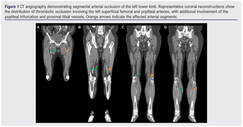
CT angiography showed extensive chronic thrombotic occlusion of the superficial femoral and popliteal arteries extending over approximately 40 cm, with additional occlusion of the popliteal trifurcation and proximal tibial vessels (Figure 1). A short intervening segment of preserved opacification suggested non-contiguous thrombosis. Minimal atherosclerotic disease was identified on imaging. A femoral-distal bypass was initially considered; however, intervention was deferred due to marked thrombocytosis (platelet count >1500×109/L) and the associated high perioperative thrombotic risk. He was admitted for optimisation of haematological parameters, including escalation of cytoreductive therapy. His regular apixaban was withheld on admission, and he was managed with low molecular weight heparin perioperatively while continuing antiplatelet therapy (aspirin).
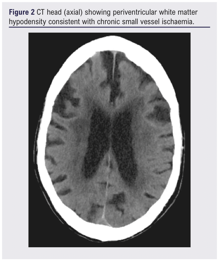
During admission he exhibited fluctuating confusion consistent with delirium on a background of cognitive impairment, impacting his ability to adhere to treatment and engage with decision-making. CT head demonstrated periventricular white matter hypodensity with mild ventricular enlargement and associated cerebral atrophy, consistent with chronic small vessel ischaemic change (Figure 2). Formal assessment showed that he lacked the capacity to make decisions regarding surgical intervention at that time. Given the urgency of his condition, with progressive limb ischaemia and tissue loss, a best-interests decision was made following discussion with his family, taking into account his previously expressed wishes to proceed with treatment.
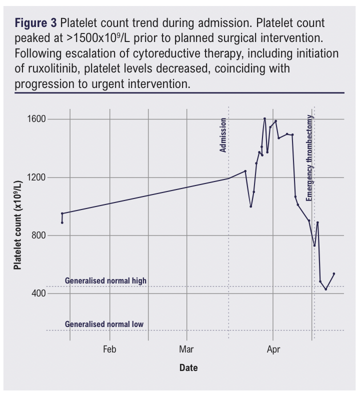
Despite medical optimisation, including escalation of hydroxycarbamide and subsequent initiation of ruxolitinib, platelet counts remained elevated (Figure 3) and his limb ischaemia progressed, with development of worsening necrosis and pain. This may have been compounded by challenges with treatment adherence in the context of delirium during admission. On 15 April 2026 his condition deteriorated to an emergency presentation with threatened limb viability.
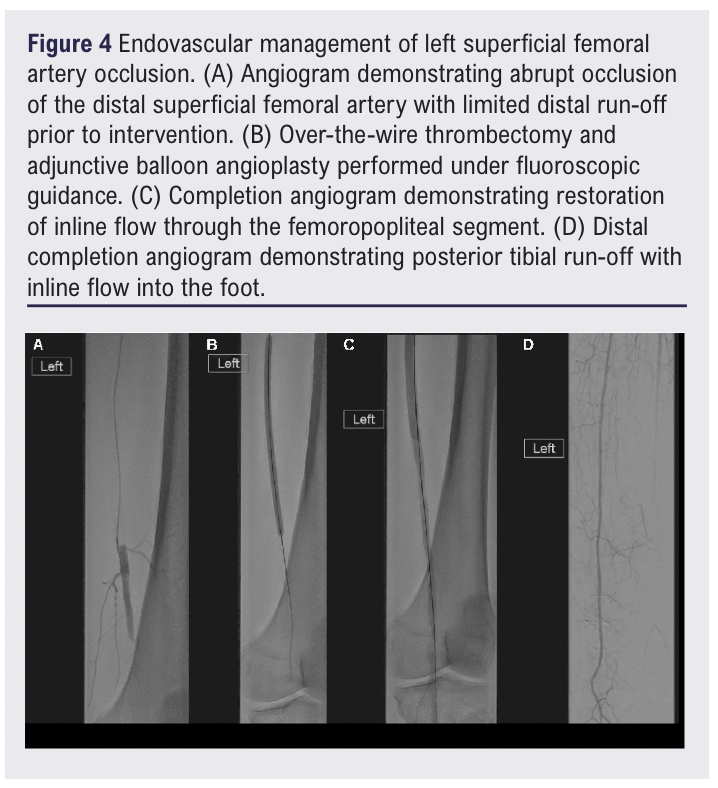
Given progressive limb-threatening ischaemia, the patient underwent urgent hybrid fluoroscopic-guided surgical thrombectomy with endovascular thrombectomy and balloon angioplasty under general anaesthesia. Following surgical exposure of the common femoral, superficial femoral and profunda femoris arteries, a transverse arteriotomy was performed in the proximal superficial femoral artery. Multiple over-the-wire thrombectomy passes were undertaken through the chronically occluded superficial femoral and popliteal arteries. Extensive organised thrombus was encountered throughout the femoropopliteal segment, with further thrombus burden extending into the distal runoff vessels. Adjunctive balloon angioplasty to break up chronic adherent thrombus throughout the arterial tree and further endovascular thrombectomy were performed. Further organised thrombus was retrieved from the distal superficial femoral, popliteal and posterior tibial circulation, with restoration of satisfactory back-bleeding. Completion angiography demonstrated restoration of inline flow to the posterior tibial artery with perfusion of the foot despite residual distal thrombus burden (Figure 4). Minimal underlying atherosclerotic disease was identified.
Postoperatively, limb perfusion immediately improved, enabling successful transmetatarsal amputation of the forefoot. In the early postoperative period the patient was clinically stable, alert and oriented, with improving symptoms and able to mobilise with physiotherapy. Doppler assessment demonstrated a return of posterior tibial arterial signals. His postoperative course was complicated by infection of the transmetatarsal amputation site, requiring further debridement and revision surgery, and by bacteraemia with Bacteroides fragilis isolated on blood cultures (18 April), for which he was treated with co-amoxiclav and metronidazole. Subsequent wound assessment demonstrated viable well-perfused tissue with improving wound edges, which subsequently healed. He was eventually ambulant and was discharged to the local community hospital for further rehabilitation before returning home.
Discussion
Peripheral arterial manifestations of polycythaemia vera are most often described in isolated case reports, with variable patterns of vascular involvement. For example, Alam et al reported a case of acute upper limb ischaemia in a young patient, characterised by thrombosis of the ulnar and digital arteries with progressive digital gangrene and autoamputation.8 In contrast, Okuda et al described acute bilateral lower limb ischaemia due to extensive large-vessel occlusion involving the common femoral, profunda femoris and popliteal arteries, requiring emergency bilateral thrombectomy.9 The present case demonstrates a distinct pattern, with segmental non-contiguous thrombosis of the femoropopliteal axis and progressive deterioration despite anticoagulation and cytoreductive therapy, ultimately necessitating urgent intervention.
Unlike previously reported cases of acute arterial thrombosis in polycythaemia vera, this case involved approximately 4 months of progressive ischaemia with chronic occlusion of more than 40 cm of the femoropopliteal axis. Extensive organised thrombus was encountered throughout the chronically occluded segment despite minimal underlying atherosclerotic disease, supporting thrombosis as the primary pathological process. Successful revascularisation was achieved using a hybrid fluoroscopic-guided surgical and endovascular thrombectomy with adjunctive angioplasty, restoring inline flow and enabling limb salvage.
A key challenge in this case was the timing of revascularisation in the context of marked thrombocytosis. Planned surgical intervention was initially deferred due to concerns regarding perioperative thrombotic risk – as available data in myeloproliferative neoplasms suggest perioperative thrombosis rates of approximately 1–7% and haemorrhagic complications of up to 8–10%, even in treated patients – and the patient was admitted for optimisation with cytoreductive therapy.13,14 However, despite escalation of treatment, platelet counts remained elevated and limb ischaemia progressed, ultimately necessitating emergency intervention. This highlights the clinical tension between delaying surgery to reduce thrombotic risk and the potential for irreversible tissue loss with ongoing ischaemia.
The platelet trend observed in this case further illustrates this dilemma. Although cytoreductive therapy led to a reduction in platelet count over time, this did not translate into clinical stability, with progression to limb-threatening ischaemia occurring during the period of attempted optimisation. Contributing factors may have included challenges with treatment adherence in the context of delirium and cognitive impairment during admission, which may reflect underlying cerebrovascular disease in a patient with a prothrombotic state. This suggests that, in selected patients, haematological control may not be achievable within a timeframe compatible with limb salvage, and that reliance on laboratory parameters alone may delay necessary intervention.
The postoperative course in this case was complicated by infection of the transmetatarsal amputation site with associated bacteraemia, necessitating additional debridement and revision surgery. This highlights the interplay between tissue viability and infection in the context of severe limb ischaemia, where residual distal thrombus and compromised microvascular perfusion may contribute to impaired wound healing. In addition, ruxolitinib therapy may have increased susceptibility to infection, as JAK inhibition has been associated with a higher incidence of infectious complications, particularly involving the skin and soft tissues.15
In this context, early revascularisation may be appropriate in selected patients with polycythaemia vera and progressive limb ischaemia, even in the presence of significant thrombocytosis. A multidisciplinary approach remains important, with decisions guided by clinical progression, limb viability and biochemical parameters. Individualised risk-benefit assessment is required in cases with evolving or multi-level arterial thrombosis.
Conclusion
This case demonstrates successful limb salvage following hybrid fluoroscopic-guided surgical and endovascular thrombectomy with adjunctive angioplasty break-up of extensive chronic femoropopliteal thrombosis in a patient with polycythaemia vera and severe thrombocytosis. Extensive organised thrombus was successfully removed despite approximately 4 months of arterial occlusion. We believe this to be the most chronically occluded limb to be salvaged with hybrid thrombectomy intervention in the published literature. Delayed haematological optimisation was associated with progression to limb-threatening ischaemia, highlighting the importance of balancing perioperative thrombotic risk against timely revascularisation.
Article DOI:
Journal Reference:
J.Vasc.Soc.G.B.Irel. 2026;Online ahead of publication
Publication date:
October 16, 2026
Author Affiliations:
1. School of Medicine and Biomedical Sciences, University of Oxford, John Radcliffe Hospital, Headington, Oxford, UK
2. Department of Vascular Surgery, Oxford University Hospitals NHS Trust, Oxford, UK
3. Department of Interventional Radiology, Churchill Hospital, Oxford University Hospitals Trust, Oxford, UK
4. Wolfson Centre for Prevention of Stroke and Dementia, Nuffield Department of Clinical Neurosciences, University of Oxford, Oxford, UK
Corresponding author:
Dominic PJ Howard
School of Medicine and Biomedical Sciences,
University of Oxford,
John Radcliffe Hospital,
Headington, Oxford
OX3 9DU, UK
Email: dominic.howard@ ndcn.ox.ac.uk